Northport Family Dental
Caring...For You.


At Northport Family Dental, we believe that we can provide you with the best possible dental care by combining old-fashioned patient-focused care with state-of-the-art treatments and technology. Our goal is to research and invest in the type of technology that we know will provide you with oral health benefits while making your treatment more efficient and comfortable.
State-of-the-Art Diagnostic Technology
Digital Intraoral X-Rays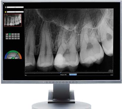
We have used digital imaging for over 20 years. Dr. Rubin was an early adopter of this technology, recognizing the direct benefit to his patients. Digital means more consistency, less exposure, and enhanced diagnostic value.
While routine x-rays are a valuable diagnostic tool, our recommendations for you are based on an individualized assessment of your needs at any given point in time. After your initial records are taken, we will often image different areas of the mouth over successive visits. This approach allows us to stay current while minimizing unnecessary films. Someone with a more extensive dental history will need to have a complete picture taken more often than someone with perfect teeth.
Digital x-rays provide a highly detailed view of your teeth, as well as other structures of your mouth. The digital sensor used to take the images mouth may resemble traditional x-ray film, but it's actually linked to a computer.
Since your x-rays are digital, we can view them together as soon as they’re taken. They are available for discussion and ultimately, for a better understanding of anything that we might need to resolve.
When you understand what’s involved, you can make better treatment decisions, which leads to a feeling of trust and confidence in your dental team.
Intraoral Camera Magnification
Intraoral cameras are a special type of technology developed specifically for use in dentistry. This handheld instrument allows us to show you images of your mouth so you get a clear picture of what’s happening and why treatment is necessary.

An intraoral camera is very small, about the size of a pen. Using this specialized equipment, we can produce highly-detailed, precise images of your teeth and gums. This information helps you see what we see and also allows us to make an accurate diagnosis.
At Northport Family Dental, our philosophy is that, if you can see it, you can understand it. And if you can see it better, you just might understand it better, too. Magnification reveals small details that help us evaluate how well the entire oral environment is or isn’t functioning. It’s easy to see a broken tooth, but it’s more helpful to recognize the underlying forces that cause the tooth to break and develop treatment strategies to prevent future problems.
The intraoral camera is an important motivational tool for children and adolescents, as well as adults. It helps them make the important connection between actions, results, and consequences. You’re also more likely to brush and floss when you understand why you need to. Patients, regardless of their age, will gain valuable insights into the condition of their oral health .
Cone Beam Computed Technology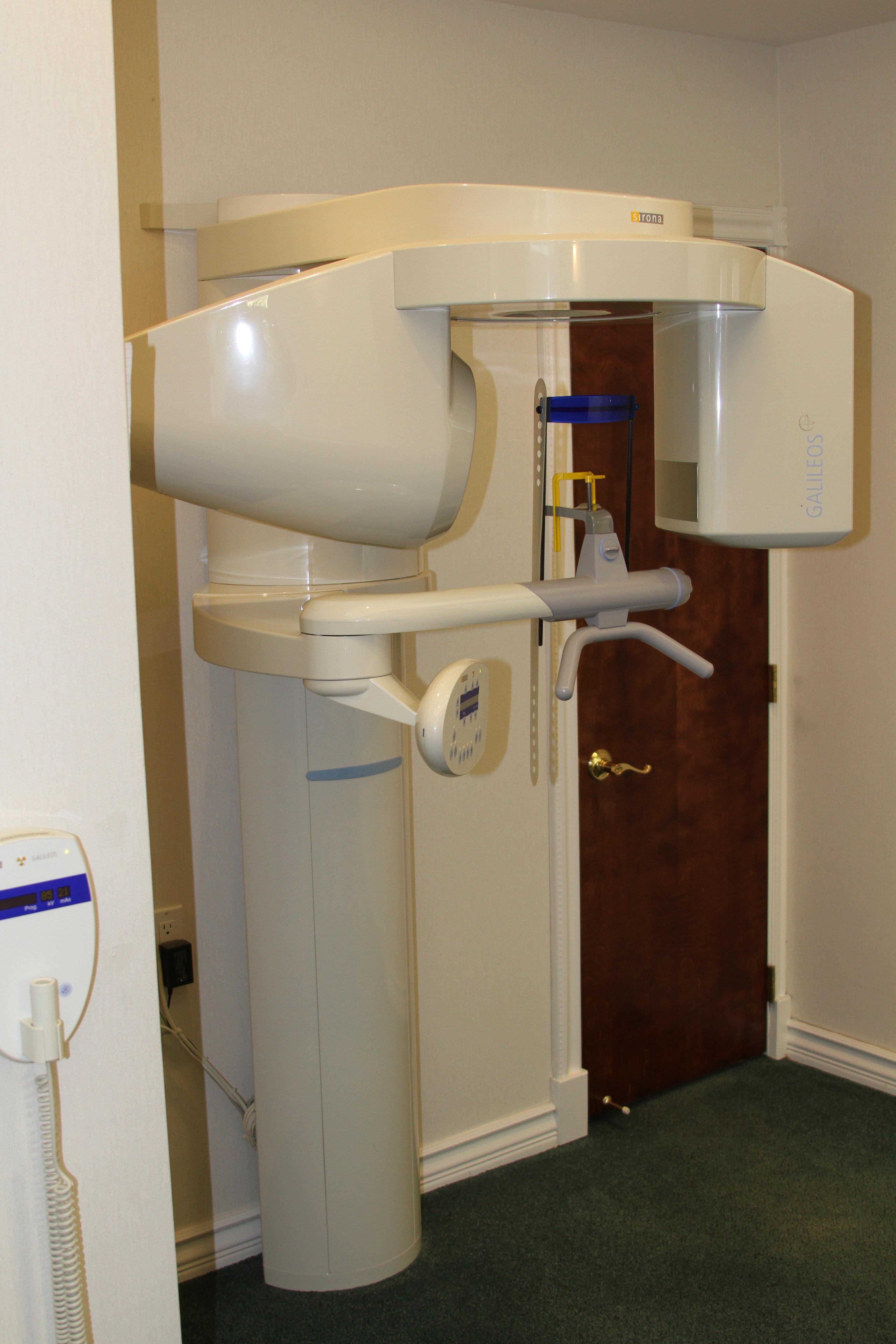
Cone beam computed tomography combines high-quality 3-D imaging with low-dose radiation. This technology projects digital x-rays in a highly accurate and controlled cone-shaped beam.
The digital x-ray scanner is mounted on a rotating arm and moves in a complete rotation around your head. Just sit back and relax and the scanner does the rest. It takes a minute or less to complete the scan, which provides us with computer-generated views of your oral and facial bone structure in 3-D.
With the level of detail provided with cone beam technology, it’s easy for us to make accurate decisions about the treatment approach that is most appropriate for your specific needs.
This technology gives us advanced diagnostic capabilities beyond the standard two-dimensional panoramic film. Our specialized software allows us to view and manipulate a 3-D reconstruction of your upper and lower jaws and teeth. This is an invaluable modality that allows us to provide our patients with the highest standard of care.
Some of its uses and benefits are:
Detection of Pathologies
Cone beam technology allows early detection of changes in the jaw related to certain disease states and conditions. Our ability to view the image from multiple angles provides a more accurate representation of size and location of the problem, which in turn helps guide our approach to the most appropriate treatment.
Implant Evaluation and Guided Surgery
At Northport Family Dental, we place the majority of our patients' implants. Viewing your jaw in 3-D allows for precise planning and more accurate placement of dental implants. This upfront planning can show us limiting factors such as inadequate volume of bone available to properly position the dental implant. For instance, the need for bone grafting prior to surgery can be identified and incorporated into the treatment plan.
Evaluation of Wisdom Teeth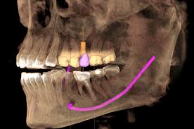
When removal of any tooth is contemplated, understanding the anatomy of adjacent teeth can be the difference between an uneventful recovery and one with unforeseen complications. An example would be paresthesia, where sensation to lower teeth and soft tissues is lost due to the severing of a nerve bundle that the roots of a wisdom tooth were wrapped around. If known ahead of time, alternative approaches can be taken and problems avoided.
Root Canal Therapy
Diseased pulp (nerve) in your tooth releases toxins into the surrounding bone. Using cone beam technology, changes in the appearance of this area can be detected earlier than with conventional x-rays. Cone beam lets us visualize the three-dimensional shape of the space we need to negotiate when we perform the root canal procedure. It helps identify additional canals, their location, and their shape. Additionally, certain fractures not visible on conventional x-ray are often seen with cone beam.
Invisalign Treatment
Adults often choose to improve their smile utilizing Invisalign aligners to move teeth. Cone beam reveals the relationships of the tooth roots to be moved during orthodontic treatment. Knowing the shapes of these roots and how adjacent structures relate to each other helps determine effective treatment.
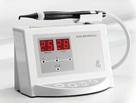
DIAGNOdent Cavity Detection
 There has been a fundamental shift over the last 50 years as to how teeth decay. Although there are many factors to be considered, the introduction of supplemental Fluoride has markedly decreased the mean number of teeth that develop decay and require treatment. Because Fluoride hardens the outer layer of protective enamel, it changes how decay progresses. Traditional tools used in dentistry are no longer adequate to detect decay in its earliest stages.
Aside from magnification, we use DIAGNOdent, a small handheld laser that objectively measures a known characteristic of enamel. It has the capability to distinguish between healthy and diseased tooth structure. A digital readout showing changes in the relative density of effected tooth structure gives us an objective measure of decay. Low numbers may indicate an area that has the potential for "healing". Monitoring the area over time to assess progression or remission now has an objective measure.
Early recognition of a developing problem area allows for less invasive solutions. Treatment can be simpler and less costly.
State-of-the-Art Equipment
CEREC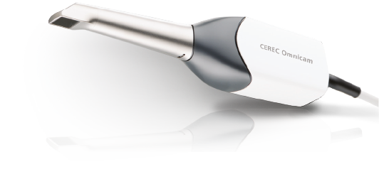
CEREC technology has transformed not only the patient experience for the better, but has allowed us to consistently provide a higher level of service with fewer visits. What used to take several appointments can now be accomplished in a single visit. As an early adopter of this technology, Dr. Rubin recognized the value of quality single-visit restorations for his patients.
Optical impressions replace traditional material filled trays that need to harden in the mouth. While this method is still utilized from time to time, most people prefer this technological advance. The majority of our Crowns, Onlays and short span Permanent Bridges are made in a single visit.
These restorations are not only strong but are life-like in appearance. Formed from a solid block of material in our milling chamber, they contain no metal. Transmitting light in a similar way as natural teeth, these materials blend into their surroundings better than traditional porcelain fused-to-metal crowns.
Computerized Porcelain Oven
Having an oven in-office expands the range of materials that can be matched to specific clinical situations. It is a necessary component in the process allowing us to provide beautiful, high quality restorations that mimic nature and withstand the test of time.
Characterization of your ceramic allows us to build in a full spectrum of color to more precisely match your adjacent teeth.
WaterLase Soft/Hard Tissue Laser
A virtual magic wand that allows us to perform many procedures more predictably, faster and with minimal need for anesthesia. The WaterLase is a water-energizing 2,780 nm YSGG laser that delivers air and water in precise proportions. to symbiotically excite water molecules from both the handpiece spray and inside the target tissue. The result is an effective biological micro-ablation of tooth structure. The atomized spray of water and air continually re-hydrates the tooth, preventing heat and pain.
Some of the procedures where we utilize this technology are:
Cosmetic Contouring of Soft Tissue
We commonly use WaterLase technology in cosmetic cases where mismatched heights of soft tissue that frame adjacent teeth result in a lack of symmetry. This often detracts from what we perceive as ideal or attractive, but a simple laser contouring is predictable, restores symmetry, and improves outcomes.
Frenectomy
The frenum is a thick or prominent muscle attachment on the upper arch between the front teeth that can result in undesirable spacing of teeth. Trying to correct this spacing includes addressing the force of the frenum, and the WaterLase simplifies this process.
You will also find a similar band of muscle under the tongue. In some children, this muscle is so short that it makes the child literally “tongue-tied.” This condition limits the ability of the tongue to extend to positions necessary for proper pronunciation.
Removal of Inflamed or Excessive Tissue in Restorative Procedures
Since the majority of our crowns and onlays are made in one visit, it’s imperative to exercise control over surrounding gum tissues and supportive bone. WaterLase allows us to contour both with minimal discomfort, less bleeding, and faster healing. Utilizing WaterLase and CEREC technology, we are able to complete these procedures in one visit.
Periodontal Treatment
WaterLase is very effective when used as an alternative to gum surgery. Studies show that laser therapy promotes regrowth of gum tissue lost due to periodontal disease. This is one of the most exciting developments in many years. Laser energy is used to reduce undesirable bacteria, clean root surfaces, and promote natural healing.
If you have difficulty using our website, please email us or call us at (631) 754-1107
View the ADA Accessibility Statement